 Possible ID: Cladosporium
Classification:
Phylum Ascomycota
Class Dothideomycete
Order Canodiales
Family Davidiellacaeae
Isolation and culturing methods:
Isolated from an agar plate that was placed near a vent in the Rooke Science at Bucknell University (Lewisburg, PA). This plate was used when we were collecting spores to differentiate from ecosystems inside and outside the lab. Plates were incubated at 20 degrees Celsius for a week or more. Then, a small volume of hyphae was excised for the agar and transferred to achieve a pure culture.
Culture appearance and growth
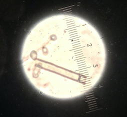
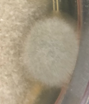
Before the was pure cultured, it formed one large circular individual. However, when it was pure cultured, it formed multiple, smaller circular individual. These looked as if they were all distinct “colonies.” It was grey and looked puffy – soft to touch. The hyphae grew out on top of it so when some was taken to pure culture, it was darker underneath. Under the microscope, I could clearly see that the hyphae were septate and around 5-6 micrometers wide
Spore production
The spores are 1-3 micrometers big. They are lightly colored. Additionally, although there are some random spores, most spores are clumped around the hypae.
Collector: Manya Group: RM2S
Culture Collection
Possible ID: Trichothecium
Classification:
Phylum Acscomycota
Class Sordariomycete
Order Hypocreales
Family Incertae sedis
Isolation and culturing methods:
Isolated from an agar plate that was used for a prior lab and was labled “TriB.” However, because it was not pure, Trichothecium grew on it. A small volume of hyphae was excised for the agar and transferred to achieve a pure culture.
Culture appearance and growth
The culture looked pink with white hyphae growing out of it. The hyphae are around 4-5 micrometers wide.
Spore production
The spores are enclosed in conidiospores around 12 microns long and 7 microns wide. The condiospores itself are septate; It looks as if it was 2 spores stuck together. This is a defining characteristic of trichothecium.
Collector: Manya Group: RM2S